Accurate quantification of cardiac anatomy and function using sparse 2D echo images, achieved with additional instrumentation during the scan and an algorithm that reconstructs the 3D shape of the heart from 2D images.
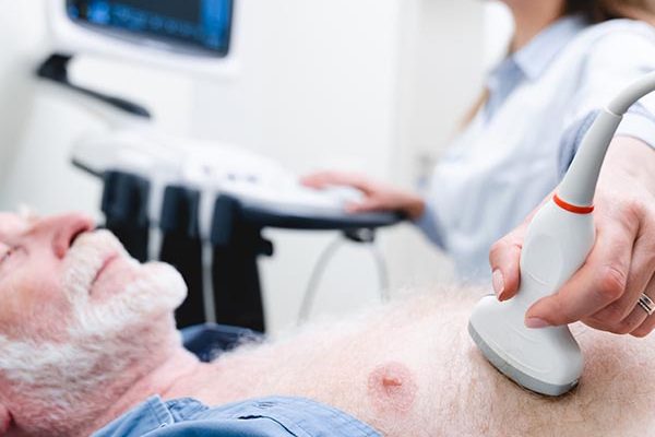
The echo images do not need to be carefully oriented and thus can be acquired by personnel with minimal training.
Proposed use
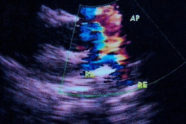
The reconstructed 3D shape of the heart over the cardiac cycle enables high quality, automated quantifications of a patient’s heart chamber structure and function, including length and thickness dimensions, volumes and stroke volumes, ejection fraction, and myocardial strains. These are important quantitative information for clinical decision-making for healthcare professionals.
Our system can be implemented in the pocket-sized, hand-held echo devices, adding low-cost and portability to its advantages. Further, since the measurement can be done by personnel with minimal training, many more clinicians can use our system, increasing access to echocardiography. This includes doctors and nurses in the emergency departments, doctors and nurses walking through wards, and clinicians in a remote, rural location. The technology can significantly shorten the waiting time for an echo scan, and allow many more scans to be conducted, improving patient care.
Problem addressed
Despite high and increasing demand, echocardiography is not always readily available to patients. This is because, traditionally, performing echocardiography requires years of specialist training and experience. Sonographers must place the transducer accurately at specific angles and positions to acquire images, otherwise there will be a lot of inaccuracies. They need knowledge and experience to interpret images when performing quantifications. For example, in the UK, patients may wait for weeks for a scan, or many hours in the emergency department, jeopardizing their health.
Our system reduces the skill level needed for echo scans and quantifications, allowing a wider range of medical personnel to conduct it and many more scans to be conducted, in many more places. This can open up access to echocardiography in scenarios that were previously difficult, and may thus open up a new and substantial market.
Because our system uses 3D reconstruction, which has precision advantages over direct quantifications from 2D images, it does not require human steps in quantifications, which also improves precision.
Technology overview
The invention combines years of expertise from clinical cardiology and Computational Bioengineering. We are seeking industry collaboration opportunities to further develop this technology to bring it to market quicker.
Benefits
- Reduce skills needed to perform echocardiography, allowing many more clinicians to perform echo, and many more times.
- Enable quantitative echo measurements using low-cost, portable point-of-care ultrasound devices, allowing echo to be performed in many more locations.
- Dramatically increase access to echocardiography, by enabling it in scenarios that are previously difficult, potentially opening up a new, substantial market
- Automatic quantifications from images removes the need for human steps, and thus reduces the associated imprecision
- Quantifications are based on 3D reconstructions, which can give precision advantages over direct measurements from 2D images.
Intellectual property information
Proprietary copyright and potential patent