More than 26 million people worldwide suffer from heart failure; there are around 1 million new cases annually in the US alone. However, most patients experience late diagnosis and poor monitoring. For this reason, heart failure commonly leads to hospitalization, has high readmission rates and has high mortality rates.
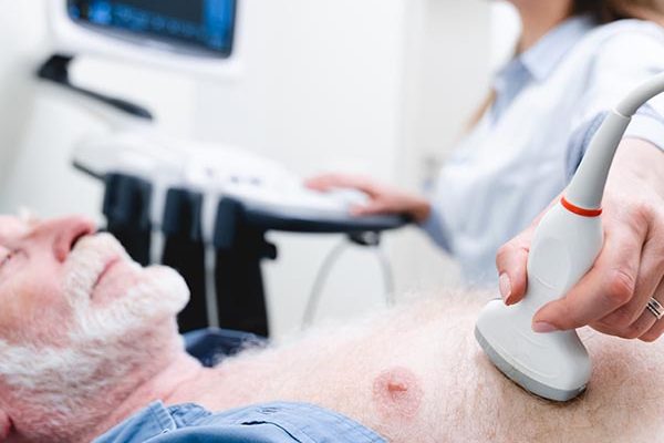
ArterioWave is a novel ultrasound-based tool for screening and monitoring those at risk of, or suffering from, heart failure. It can be used by non-specialists. It can be implemented on ultrafast scanners, conventional scanners or handheld devices.
Proposed use
- Screening and disease management
- Point of care testing
- Treatment decision support
Problem addressed
People developing heart failure typically present to their GP with non-specific symptoms. For a definitive diagnosis, they should be referred to a specialist centre for echocardiography. However, 79% of patients are diagnosed after an emergency hospital admission, even though 41% had visited their GP with symptoms in the preceding 5 years (source: BHF). Patients seldom receive more than one echocardiogram. Monitoring disease progression, titrating medication, assessing compliance and discharging patients from hospital rely on subjective assessment.
Previous studies have shown that diagnosis and prognosis can be achieved by analysing waves in arteries. However, current methods of Wave Intensity Analysis are invasive, inaccurate or expensive. ArterioWave is non-invasive, accurate, inexpensive and simple to use, and will allow GPs, sonographers and nurses, as well as specialists, to offer patients earlier, easier, and more frequent evaluation of heart function in the community and on the ward.
Technology overview
When the left ventricle contracts, a wave of increased blood pressure, blood velocity and arterial diameter propagates along the systemic arteries. When the ventricle relaxes, a wave of decreased blood pressure, blood velocity and arterial diameter occurs. Six previous trials have shown that wave intensities can indicate the severity of heart failure, predict outcomes, and discriminate between heart failure with or without preserved ejection fraction.
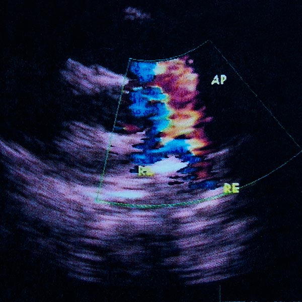
ArterioWave senses changes in wave intensity continuously over the cardiac cycle from spatially and temporally coincident measurements of blood velocity and arterial diameter. Velocity and diameter can be measured using B-mode or M-mode ultrasound:
- B-mode / ultrafast ultrasound: Ultrasound Image Velocimetry in conjunction with plane-wave ultrafast ultrasound gives arterial blood velocity and diameter from the same images, without contrast agent. (Pre-clinical trials completed; in patient trial)
- M-mode / conventional or PoC ultrasound: A single ultrasound beam is fired across the artery twice in quick succession. Diameter is obtained by standard methods and velocity by correlating the pattern of scatterers along the beam at the two times. (In development)
Benefits
- Clinically proven parameters: Analyses of wave intensity indices that closely relate to heart failure in human trials
- Simple to use: Measures the function of heart through any arterial pulse accessible to ultrasound
- Easy data interpretation: Intuitive readout. No ultrasound image interpretation required
- Safe: Risk-free even for frequent use on patients with a high heart failure risk
- Maximised Compliance: A truly non-invasive technique that does not require catheter insertion or contrast agent
Intellectual property information
GB2570131A1 FLUID FLOW ANALYSIS
WO 2019138241 A1 FLUID FLOW ANALYSIS
US 2019099153 A1 FLUID FLOW ANALYSIS
EP3432802 B1 FLUID FLOW ANALYSIS (France, Germany, Netherlands, UK)
CN 109152563 A 流体流分析
Canada patent pending


