Technology Overview
This system combines imaging biomarkers and automated image processing to diagnose fertility pathologies in male and female reproductive organs, making use of super resolution ultrasound localisation microscopy (ULM) using microbubble contrast enhancement. In this system, image processing and biomarker generation are automated, making the technology simple to use and providing a pathway to easy integration into clinical ultrasound scanning hardware.
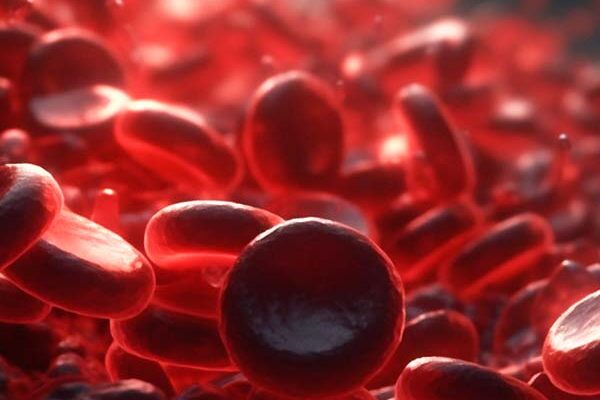
ULM is an advanced type of ultrasound imaging that tracks the movement of injected microbubbles over time to examine microvessels or microvasculature – the tiny blood vessels found within all organs and they are crucial for supplying oxygen and nutrients to tissues. This makes imaging of microvessels a highly sensitive method of assessing the health and function of any given tissue.
Problem addressed
Infertility affects 15% of couples trying to conceive, globally, with causative abnormalities distributed evenly among men and women. Furthermore, over 90% of young women experience pain or bleeding requiring medical assessment. Commonly used first-line investigations for these conditions are time- and resource-intensive and potentially embarrassing for patients.
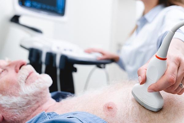
Ultrasonography is increasingly being used in clinical practice to confirm one of several suspect diagnoses known to manifest in male and female infertility. The reproductive organs are ideal for ULM because they are close to a surface, such that a hand-held ultrasound probe can be used. Reproductive organ pathologies are also commonly associated with abnormalities in microvessel characteristics.
Proposed Use
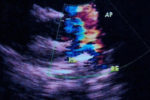
ULM and advanced image processing can measure, with superior precision, multiple parameters relevant to the diagnosis of indications related to vascular disease. These include microvessel density, microvascular flow, regularity of the flow, and tortuosity of the vessels.
In the male reproductive system, this technology has been shown to detect hypogonadism (testosterone deficiency) and male infertility based on dysfunction of the testes. It may also be used to guide testicular sperm extraction in men with azoospermia.
In the female reproductive system, this may be applied to diagnosing menorrhagia, endometriosis, endometrial hyperplasia, gestational trophoblastic disease. It may also be used in troubleshooting the failure of assisted reproductive technology treatments and adverse pregnancy outcomes.
This system is also being explored in the detection and mapping of cancers in the reproductive system for both men and women.
This technique holds novelty over other reproductive diagnostic tools currently on the market, as there are no current imaging solutions for azoospermia, hypogonadism, or endometriosis. As well as no accurate approaches to predict the poor implantation of embryos into the endometrium following fertility treatment.
Benefits
- Compatibility – this technology is software-based and is able to merge with existing ULM systems
- Versatile application – use in women’s and men’s reproductive health, targeting various conditions
- Breakthrough – screening for conditions where there are no current imaging diagnostics
- Non-invasive and time-efficient – reduces patient discomfort during diagnosis
- Efficient operation – advanced signal processing provides quality results through improved signal-to-noise ratio
Intellectual property information
PCT Application – WO2024161154A1 – Treatment of male reproductive disorder
Software and knowhow IP has also been developed for the purpose of imaging the microvasculature of the endometrium and ovaries.
Inventor information
Dr Channa Jayasena – Channa is an internationally recognized specialist in the field of reproductive endocrinology. He currently leads a research team at Imperial College London and works as Consultant in Reproductive Endocrinology and Head of Andrology at St. Mary’s & Hammersmith Hospitals in London. His Imperial-led invention successfully delivered the MRC-industry collaboration with Astra Zeneca, producing US FDA-approved neurokinin 3 inhibitors for menopause. He is widely featured in media on healthcare advances, including BBC, The Economist, CNN, Guardian, The Times, and The Daily Mail.
Professor Jipeng Yan – Jipeng is a Professor at the School of Mechatronics Engineering and the State Key Laboratory of Robotics and Systems at Harbin Institute of Technology, China, where his research concerns the use of SRUS/ULM medical ultrasound imaging and the utilization of robotics for ultrasound to further improve imaging quality and reliability. While at Imperial, Jipeng was a Chan-Zuckerberg Initiative-funded Research Associate in the Ultrasound Lab for Imaging and Sensing (ULIS) lab.
Professor Mengxing Tang – Mengxing is a Professor of Biomedical Imaging in Imperial College’s Department of Bioengineering and Principal Investigator in the ULIS lab. His research mainly focuses on developing new in vivo imaging techniques of ultrahigh temporal resolution and spatial resolution using ultrasound, signal and image processing techniques, for visualising and quantifying macro- and micro-vascular structure and flow dynamics.
Professor Adrian Lim – Adrian is a Professor of Practice (Radiology) at Imperial College’s Department of Metabolism, Digestion, and Reproduction, consultant radiologist at Imperial College Healthcare NHS Trust, and Head of Ultrasound at Charing Cross Hospital. His specialist areas include advanced clinical applications of ultrasound and translational use of novel techniques in functionally assessing disease processes.
Professor Suks Minhas – Suks is a consultant andrologist at Imperial College Healthcare NHS Trust and is the centre director of the European Academy of Andrology. His fields of expertise include sperm retrieval techniques including micro TESE, Peyronie’s disease surgery, penile reconstruction, testicular cancer and testis sparing surgery, erectile dysfunction and penile implant surgery.