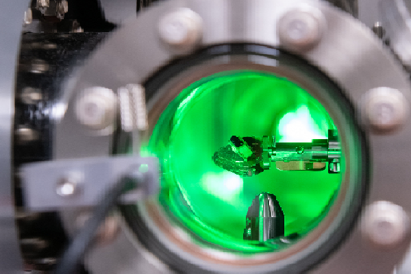
A fast, achromatic, and light-efficient optical system that can record super-resolved Structured Illumination Microscopy (SIM) images at very high speeds, using only a single moving part. This setup offers a cutting-edge tool for researchers seeking precise insights into dynamic cellular activities at the nanoscale.
Proposed use
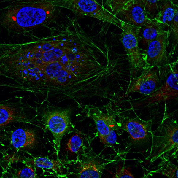
This novel SIM system facilitates the real-time observation of fast micro-biological phenomena with spatial features smaller than the diffraction limit of light (down to 120 nm), and with a remarkably-high temporal resolution (currently as short as 35ms per super-resolved frame, but with scope for improvement). This advancement is particularly advantageous for studying cell structures and mechanisms, such as observing endocytosis in Plasmodium malaria, monitoring calcium activity in dendritic spines, and examining the growth of actin micro-filaments.
Problem addressed
SIM enables multi-color live cell imaging at double the resolution of conventional microscopes. Despite its advantages, existing commercial systems, such as the Nikon N-SIM S, Zeiss Elyra 7, and GE Healthcare Deltavision OMX, face several critical issues including high price, complex design and slow imaging speed. This invention significantly improves the affordability, speed and overall performance of SIM by introduction of a simplified hardware design that requires only one moving part.
Technology overview
This invention allows users to create arbitrary fringe patterns and switch between them at high speed. The system can be demonstrated on a SIM system, creating a fast, achromatic and light-efficient system that can record super-resolved images at very high speeds.
Benefits
- Simplified hardware redesign for SIM, using only a single moving part
- High frame rate for 2D and 3D imaging (52 frames/second and 31 frames/second respectively)
- Achromatic (adaptable to multicolour SIM)
- Cheaper and easier to build / maintain
- Scope for further cost and size reduction
Intellectual property information
2306130.2/GB/PRV


