Surgical Polarimetric Endoscopy (SPE) Device

Reference number 7005

Sectors: Medtech

Industries: Medical Devices, Medical Imaging, Medical Technology

A novel endoscopic device for the acquisition of high resolution, polarization-resolved images of tissue allowing improved cancer detection

Proposed Use

  • This surgical device is intended for explorative endoscopy procedures for pathological tissue examination.
  • The device may be applied in laryngoscopy (or arthroscopy/ laparoscopic) procedures

Problem Addressed

Surgical endoscopy plays a key role in laryngeal cancer diagnosis and intervention. Currently, surgeons rely on standard white light endoscopy (WLE) to detect laryngeal pathologies based on colour contrast, which can lead to unsatisfactory sensitivity and high miss rates. There is a pressing need for a system that enables enhanced tissue contrast beyond standard colour appearance to include the polarization properties of healthy and pathological tissues to improve the rate of cancer detection.

Technology Overview

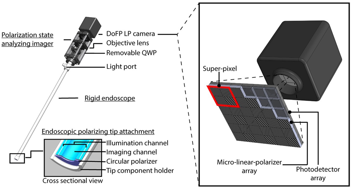
The system relies on a modification of the illumination path and camera detection system and consists of four different modules:

  1. a rigid endoscope with in-built imaging and illumination channels
  2. an endoscopic tip attachment holding a ring-shaped circularly polarizing plate over the illumination channel at the distal end of the endoscope
  3. a polarizing state analysing imager consisting of a division of focal plane linear polarimetric (DoFP-LP) camera
  4. an endoscope light source

A breakdown of the construction of the SPE device. The modules that form the device are described in the text before this image. The endoscopic tip attachment contains an imaging channel surrounded by an illumination channel, surrounded by a circular polarizer and contained within a tip component holder. The DoFP LP camera is shown to consist of a micro-linear-polarizer array and a photodetector array

Benefits

  • The first in vivo intraoperative polarimetric endoscope
  • Lightweight and compact design
  • Real time, label-free analysis
  • Wide field, high resolution imaging
  • Enhanced imaging capabilities to improve tumor detection and cancer diagnosis

Developmental Stage

  • The device is ready for demonstration
  • The technology requires further clinical testing
  • TRL 4 to 5

Intellectual property information

International PCT patent application pending

Inventor

Professor Daniel Elson

Professor of Surgical Imaging
Faculty of Medicine, Department of Surgery & Cancer

Visit personal site

Contact us about this technology


Contact

Dr Louie Scott

Industry Partnerships and Commercialisation Officer, Faculty of Medicine

Louie is Industry Partnerships and Commercialisation Officer within Imperial Enterprise, supporting inventions arising from the Faculty of Medicine

Contact Louie

+44 7517 551871

[email protected]

Related technologies

A technique to detect and discriminate mycobacteria using intact cell lipidomics

A technique to detect and discriminate mycobacteria using intact cell lipidomics

A technique to detect and discriminate mycobacteria using intact cell lipidomics Find out more

A Wearable Echolocation Device for the Visually Impaired

A Wearable Echolocation Device for the Visually Impaired

Wearable enabling visually impaired user to hear echoes bounced back from structures and objects, allowing the user to perceive their spatial environment Find out more

Data-driven Multiplexing for Accurate Gene Detection

Data-driven Multiplexing for Accurate Gene Detection

Machine learning approach for high-level multiplexing in qPCR and dPCR, up to 21-plex demonstrated Find out more

Sign up for updates

Sign up for monthly technology alerts via email, and find other ways to connect with us.

Loading...