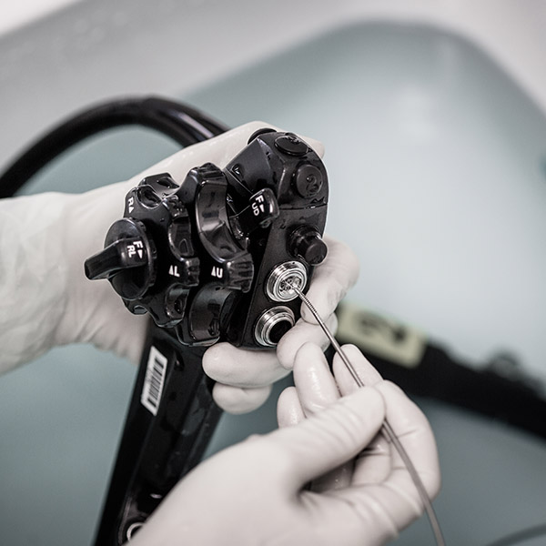
EndoDrone is a novel robotic optical biopsy scanning framework designed to improve the sensitivity of gastrointestinal (GI) endoscopy by automated scanning and real-time classification of wide tissue areas based on optical data. A “hot-spot” map is generated to highlight dysplastic or cancerous lesions for further scrutiny or concurrent resection. The device uses hyperspectral (HS) optical biopsy and works as an accessory to a conventional endoscope. The device is compatible with the anatomical dimensions of the colon and the oesophagus.
Proposed Use
An exploratory device for improving cancer detection rates, especially for small flat dysplastic lesions (<5mm). EndoDrone can be employed as a standalone device or in combination with other optical and non-optical single-point probe modalities (fluorescence, fluorescence lifetime, polarised, confocal, Raman, optical coherence tomography, ultrasound).
Problem Addressed
GI endoscopy is the gold-standard procedure for detection and treatment of dysplastic lesions and early-stage cancers. Despite its proven effectiveness, its sensitivity remains suboptimal due to the subjective nature of the examination, which is highly reliant on human operator skills and expertise. For bowel cancer, colonoscopy can miss up to 22% of dysplastic lesions, with even higher miss rates for small (<5 mm diameter) and flat lesions.
Technology Overview
The key features of this technology are:
- 2D and 3D reconstructions of endoscopic HS optical biopsy data acquired via robotic scanning by a radial array of contact single-point optical probes
- Sub-millimetre optical resolution and automated HS image segmentation, to allow the identification of frequently missed flat pre-cancerous lesions
- In vitro studies showed that the smallest simulated flat pre-cancerous lesions resolved by the device were 0.75 mm in diameter
- The size of the device is compatible with the anatomical dimensions of the colon and oesophagus
- The device does not compromise the use of the flexible endoscope’s working channels, avoiding workflow interference
Benefits
- Rich diagnostic optical biopsy data
- Real-time automated image segmentation allowing the generation of a “hot-spot” map
- Compatible size with the anatomical dimensions of the colon and oesophagus
- Improved detection rate of small flat dysplastic lesions (<5mm)
Development Stage
- Device validated in vitro
- Preliminary animal studies performed
- Exploring a miniaturised version and applications to other non-GI hollow organs (e,g., upper airway, bronchi, bladder)
- TRL 3-4
Intellectual Property
Granted patent in Japan (JP6634548B) and published patent applications in Europe (EP3264967A1) and the US (US2018042457A1)


